
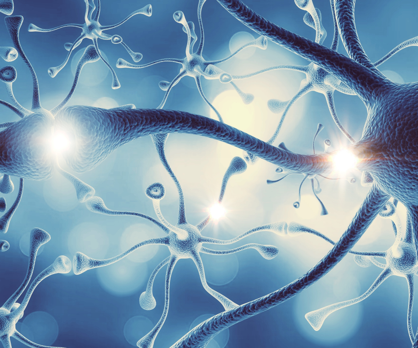
Peripheral Nervous System

Peripheral Nervous System: Understanding Its Structure and Function

The Peripheral Nervous System (PNS) is the communication network that connects the brain and spinal cord to the rest of the body. It plays a vital role in movement, sensation, and the regulation of many automatic body functions.

Without the Peripheral Nervous System, the brain would not receive information from the body, and muscles would not receive instructions to move. Every voluntary movement, reflex, and sensory experience depends on this complex network of nerves.

Understanding the Peripheral Nervous System is essential for fitness professionals, healthcare providers, and anyone interested in human anatomy and physiology.

What Is the Peripheral Nervous System?

The Peripheral Nervous System includes every nerve located outside the brain and spinal cord. While the central nervous system (CNS) processes information, the PNS serves as the communication pathway between the CNS and the rest of the body.

The PNS collects sensory information from muscles, skin, joints, and internal organs. It then carries this information to the brain and spinal cord for processing. At the same time, it delivers motor commands from the CNS to muscles and glands.

This constant exchange of information allows the body to respond quickly and efficiently to internal and external changes.

Main Functions of the Peripheral Nervous System

The Peripheral Nervous System performs several essential functions.

These include:

  • Carrying sensory information to the central nervous system.
  • Delivering motor commands to skeletal muscles.
  • Regulating involuntary body functions.
  • Supporting reflex actions.
  • Helping maintain homeostasis.

Without these functions, everyday activities such as walking, talking, and maintaining balance would not be possible.

The Two Main Divisions of the Peripheral Nervous System

The Peripheral Nervous System is divided into two primary sections.

Somatic Nervous System

The somatic nervous system controls voluntary movement and conscious sensation.

It allows individuals to intentionally move skeletal muscles and perceive sensations such as touch, temperature, pressure, and pain.

Examples include:

  • Walking.
  • Lifting weights.
  • Typing on a keyboard.
  • Feeling a hot surface.

The somatic nervous system communicates directly with skeletal muscles through motor neurons.

Autonomic Nervous System

The autonomic nervous system (ANS) regulates involuntary body functions.

These activities occur automatically without conscious thought.

Examples include:

  • Heart rate.
  • Blood pressure.
  • Digestion.
  • Breathing rate.
  • Pupil size.

The autonomic nervous system helps maintain the body’s internal balance throughout the day.

Divisions of the Autonomic Nervous System

The autonomic nervous system has two major branches.

Sympathetic Nervous System

The sympathetic nervous system prepares the body for stressful situations.

This response is commonly called the “fight-or-flight” response.

During activation:

  • Heart rate increases.
  • Blood pressure rises.
  • Pupils dilate.
  • Blood flow shifts toward working muscles.
  • Digestion temporarily slows.

These changes help the body respond quickly to physical or emotional stress.

Parasympathetic Nervous System

The parasympathetic nervous system promotes relaxation and recovery.

It is often called the “rest-and-digest” system.

During activation:

  • Heart rate slows.
  • Digestion increases.
  • Energy stores are replenished.
  • The body conserves energy.

Both branches continuously work together to maintain balance.

Sensory Neurons

Sensory neurons, also called afferent neurons, carry information from receptors throughout the body to the brain and spinal cord.

They detect many different types of stimuli.

These include:

  • Touch.
  • Temperature.
  • Pain.
  • Pressure.
  • Vibration.
  • Joint position.

This information helps the CNS understand what is happening both inside and outside the body.

Motor Neurons

Motor neurons, also known as efferent neurons, transmit commands from the central nervous system to muscles and glands.

They allow the body to perform voluntary movements such as running, jumping, or lifting an object.

Motor neurons also control involuntary muscle activity through the autonomic nervous system.

Cranial and Spinal Nerves

The PERIPHERAL NERVOUS SYSTEM contains two major groups of nerves.

Cranial Nerves

There are 12 pairs of cranial nerves.

These nerves originate directly from the brain.

They control functions involving the head, face, neck, and several internal organs.

Examples include vision, hearing, facial movement, taste, and swallowing.

Spinal Nerves

There are 31 pairs of spinal nerves.

These nerves emerge from the spinal cord.

They carry sensory and motor information between the CNS and the trunk, arms, and legs.

Spinal nerves are responsible for most movement and sensation throughout the body.

Reflex Actions

One of the fastest responses controlled by the PERIPHERAL NERVOUS SYSTEM is the reflex.

A reflex is an automatic response to a stimulus.

It occurs without conscious thought.

For example, touching a hot surface causes the hand to pull away almost immediately.

This rapid response protects the body from injury.

Although the brain becomes aware of the event shortly afterward, the spinal cord coordinates the initial reflex.

The Peripheral Nervous System and Exercise

Exercise relies heavily on the Peripheral Nervous System.

Every movement begins with nerve signals traveling from the brain through motor neurons to skeletal muscles.

As muscles contract, sensory receptors continuously send information back to the CNS.

This feedback allows the body to adjust movement, maintain posture, and improve coordination.

During resistance training, repeated practice strengthens communication between the nervous system and muscles.

This process contributes to improved neuromuscular coordination and greater movement efficiency.

Common Disorders of the Peripheral Nervous System

Damage to the Peripheral Nervous System can affect movement, sensation, or both.

Common conditions include:

  • Peripheral neuropathy.
  • Carpal tunnel syndrome.
  • Sciatica.
  • Bell’s palsy.
  • Nerve compression injuries.

Symptoms may include:

  • Numbness.
  • Tingling.
  • Weakness.
  • Burning sensations.
  • Reduced coordination.

Treatment depends on the underlying cause and may involve physical therapy, medication, lifestyle changes, or surgery.

Why the Peripheral Nervous System Matters

The Peripheral Nervous System allows the brain and spinal cord to communicate with every part of the body. It makes voluntary movement possible, regulates automatic body functions, and provides continuous sensory feedback.

Whether walking, exercising, maintaining balance, or reacting to danger, the PNS is constantly transmitting information that keeps the body functioning efficiently.

For fitness professionals, understanding the Peripheral Nervous System helps explain movement, coordination, motor control, and exercise performance. It also provides a foundation for recognizing how nerve function influences strength, balance, and overall physical health.
Biomechanics

Biomechanics: Understanding Human Movement

Biomechanics is the study of how forces interact with the body during movement. It combines principles of anatomy, physics, and physiology to explain how and why the human body moves.

Understanding biomechanics helps fitness professionals, coaches, and healthcare providers analyze movement patterns, improve performance, and reduce injury risk.

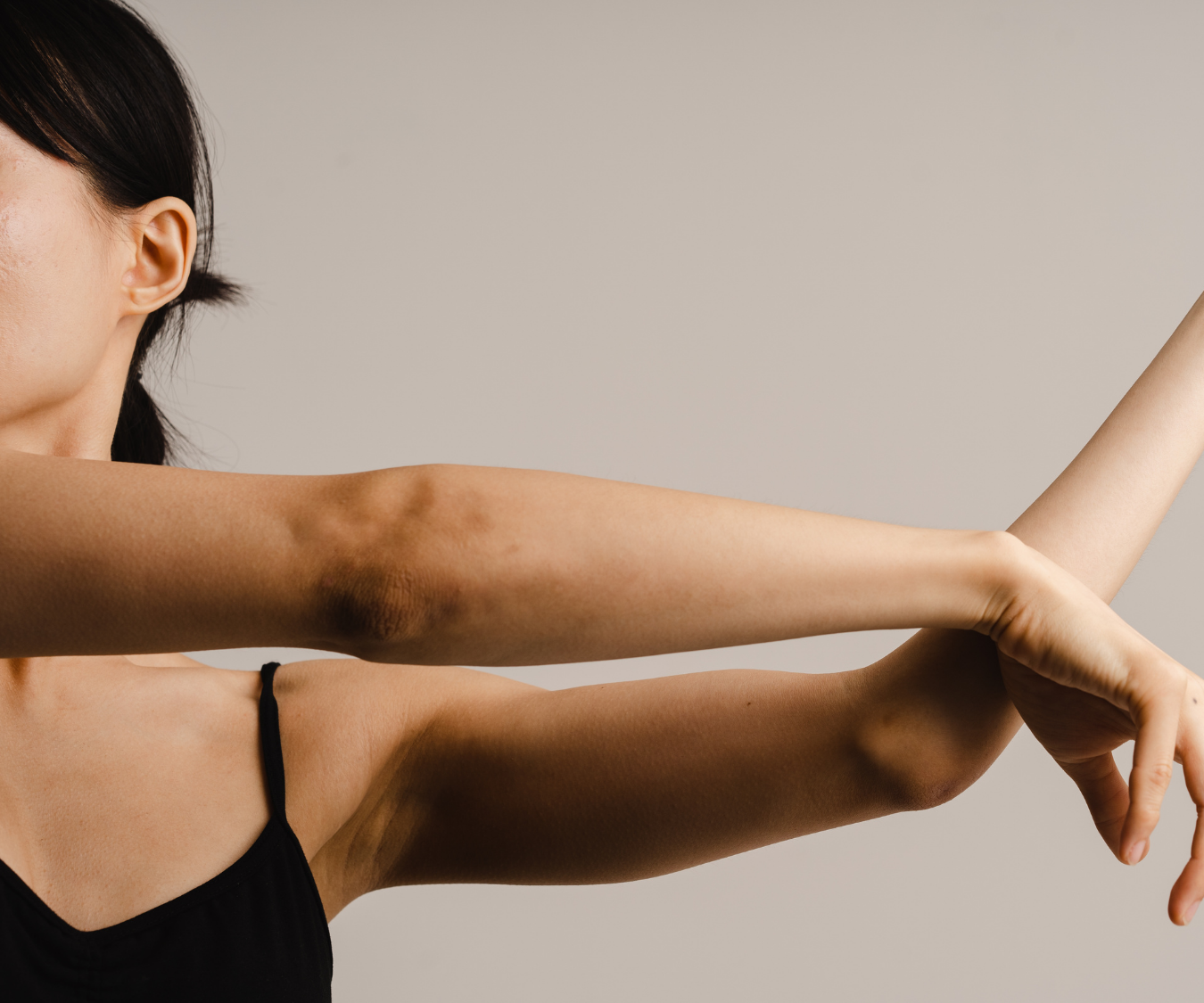
The body moves through specific anatomical planes and uses specific movement terms to describe the direction and action occurring at a joint.

Anatomical Position and Movement

Most movement descriptions begin from the anatomical position. This is when the body is standing upright, facing forward, with arms at the sides and palms facing forward.

Movement terms describe how body parts change position relative to this starting point.


Terms for Anatomical Movement

Abduction

Definition: Movement away from the midline of the body.

Example:
Raising the arms out to the sides during a lateral raise.


Adduction

Definition: Movement toward the midline of the body.

Example:
Lowering the arms back toward the body after a lateral raise.


Flexion

Definition: Movement that decreases the angle between two body parts.

Example:
Bending the elbow during a biceps curl.


Extension

Definition: Movement that increases the angle between two body parts.

Example:
Straightening the elbow after a biceps curl.


Lateral Flexion

Definition: Flexion occurring in the frontal plane.

Example:
Bending the torso sideways toward the right or left.


Scapular Movements

The scapula (shoulder blade) has unique movements that are important for upper-body function.

Protraction

Definition: Abduction of the scapula.

Example:
Moving the shoulder blades forward, such as during a push-up.


Retraction

Definition: Adduction of the scapula.

Example:
Pulling the shoulder blades together during a rowing movement.


Elevation

Definition: Movement in a superior direction.

Example:
Shrugging the shoulders upward.


Depression

Definition: Movement in an inferior direction.

Example:
Moving the shoulders downward after a shrug.


Foot and Ankle Movements

The ankle and foot have specialized movements that support balance, walking, and athletic performance.

Plantar Flexion

Definition: Extension of the foot downward (inferiorly).

Example:
Pointing the toes downward like pressing a gas pedal.


Dorsiflexion

Definition: Flexion of the foot upward (superiorly).

Example:
Lifting the toes toward the shin.


Inversion

Definition: Movement of the sole of the foot toward the median plane.

Example:
Rolling the bottom of the foot inward.


Eversion

Definition: Movement of the sole of the foot away from the median plane.

Example:
Rolling the bottom of the foot outward.


Rotational Movements

Rotation occurs when a body part turns around its axis.

External Rotation

Definition: Rotational movement away from the midline.

Example:
Turning the shoulder outward during a movement.


Internal Rotation

Definition: Rotational movement toward the midline.

Example:
Turning the shoulder inward.


Complex Joint Movements

Circumduction

Definition: Circular movement of a limb extending from the joint where the movement is controlled.

Example:
Making a circular motion with the arm at the shoulder joint.

Circumduction combines multiple movements, including:

  • Flexion
  • Extension
  • Abduction
  • Adduction

Forearm and Hand Movements

Pronation

Definition: Turning the palm or arch of the foot downward.

Example:
Rotating the forearm so the palm faces the floor.


Supination

Definition: Turning the palm or arch of the foot upward.

Example:
Rotating the forearm so the palm faces upward.


Movement Beyond Normal Range

Hyperextension

Definition: A position that extends beyond anatomical neutral.

Example:
Extending the neck or spine past its normal alignment.


Directional Movement Terms

Ipsilateral

Definition: Movement occurring on the same side of the body.

Example:
The right arm and right leg moving together.


Contralateral

Definition: Movement occurring on opposite sides of the body.

Example:
The right arm and left leg moving together during walking.


Lateral

Definition: Situated away from the midline.

Example:
The ears are lateral to the nose.


Medial

Definition: Situated toward or closer to the midline.

Example:
The nose is medial to the ears.


Why Understanding Biomechanics Matters

Biomechanics provides the foundation for understanding human movement. Knowing anatomical movement terms allows professionals to describe exercises accurately, identify movement limitations, and improve training programs.

For example:

  • A squat involves hip and knee flexion during the lowering phase.
  • A push-up involves elbow flexion and extension.
  • A shoulder press involves shoulder abduction and elbow extension.

By understanding how forces interact with the body, movement professionals can create safer and more effective exercise programs. Biomechanics helps explain not only what the body does, but also why it moves the way it does.

Skeletal Muscle

Skeletal Muscle Fiber Arrangement and Its Role in Human Movement

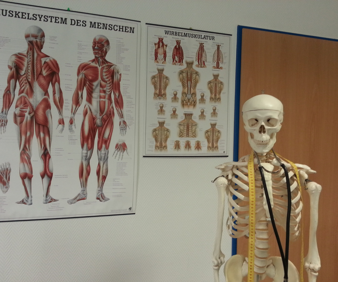
The human body relies on muscles to create movement, maintain posture, and generate force. Among the different types of muscle tissue, skeletal muscle plays the primary role in voluntary movement. It allows individuals to walk, run, lift objects, maintain balance, and perform daily activities.

The organization of skeletal muscle fibers directly influences how muscles produce force. The direction, arrangement, and structure of muscle fibers determine the amount of strength a muscle can create, the distance it can move a joint, and the type of movement it can produce.

Understanding muscle fiber arrangement is important for exercise professionals because it helps explain why different muscles perform different functions and why certain exercises target muscles in specific ways.

What Is Skeletal Muscle?

Skeletal muscle is a type of muscle tissue attached to bones by tendons. When skeletal muscle contracts, it pulls on bones and creates movement at joints.

Unlike smooth muscle and cardiac muscle, skeletal muscle is under voluntary control. This means individuals can consciously activate these muscles to perform movements.

Skeletal muscle has several important functions:

  • Producing body movement
  • Maintaining posture
  • Stabilizing joints
  • Generating heat
  • Supporting physical performance

Each skeletal muscle is made up of thousands of muscle fibers organized into bundles. The arrangement of these fibers affects how efficiently the muscle can produce force.

Muscle Fiber Direction and Movement

The direction of muscle fibers determines how a muscle pulls on a bone.

Muscles generally produce force by shortening along the direction of their fibers. Because of this, fiber arrangement influences the type of movement a muscle can create.

For example:

  • Fibers arranged in a straight line typically allow greater range of motion.
  • Fibers arranged at an angle often allow greater force production.
  • Multiple fiber arrangements create different movement abilities.

The structure of a muscle provides clues about its function.

Parallel Muscle Fiber Arrangement

In a parallel arrangement, muscle fibers run along the same direction as the length of the muscle.

These muscles are designed for greater movement distance and speed.

Because fibers shorten along their entire length, parallel muscles can create large ranges of motion.

Examples include:

  • Sartorius muscle
  • Rectus abdominis

Parallel muscles are often effective for movements requiring flexibility and longer motion.

However, they may produce less force compared with muscles that have more densely packed fibers.

Fusiform Muscle Arrangement

A fusiform muscle is a type of parallel arrangement where the muscle has a wider middle section and narrower ends.

The fibers run parallel to the muscle’s long axis.

This design allows the muscle to shorten significantly during contraction.

An example of a fusiform muscle is the biceps brachii.

During a biceps curl, the fibers shorten and pull on the forearm to create elbow flexion.

Pennate Muscle Fiber Arrangement

Pennate muscles have fibers that attach at an angle to a central tendon.

This arrangement allows more fibers to fit into a smaller space.

Because more fibers can contract at the same time, pennate muscles can produce greater force.

The tradeoff is that they usually have a smaller range of motion.

There are three main types of pennate arrangements:

  • Unipennate
  • Bipennate
  • Multipennate

Unipennate Muscles

In unipennate muscles, fibers attach to only one side of a tendon.

This design allows a large number of fibers to contribute to force production.

An example is the extensor digitorum muscle in the forearm.

Bipennate Muscles

Bipennate muscles have fibers attached to both sides of a central tendon.

This arrangement increases the number of muscle fibers that can fit into the muscle.

The increased fiber density allows greater force production.

The rectus femoris muscle of the quadriceps is an example of a bipennate muscle.

Multipennate Muscles

Multipennate muscles contain multiple sections of fibers arranged around several tendons.

This structure allows very high force production.

An example is the deltoid muscle of the shoulder.

The deltoid helps create shoulder movement in multiple directions because of its complex fiber arrangement.

Circular Muscle Arrangement

Circular muscles have fibers arranged in a circular pattern around an opening or passage.

When these muscles contract, they decrease the size of the opening.

These muscles are also called sphincter muscles.

Examples include:

  • Orbicularis oculi around the eye
  • Orbicularis oris around the mouth

Their primary function is controlling movement through openings.

How Fiber Arrangement Affects Strength

The arrangement of skeletal muscle fibers directly affects force production.

Muscles with more fibers packed into a specific area generally produce more force.

Pennate muscles often generate higher levels of force because their angled fibers allow more contractile tissue to attach to tendons.

However, increased force often comes with reduced shortening distance.

This creates a balance between strength and range of motion.

How Fiber Arrangement Affects Range of Motion

Muscles with fibers arranged parallel to their length can usually shorten more.

This creates greater movement distance.

For example, muscles involved in large movements often require greater range of motion.

Pennate muscles, while stronger, typically create less movement because their fibers do not shorten the entire length of the muscle.

The body uses different muscle designs to meet different movement demands.

Skeletal Muscle and Exercise Training

Exercise can influence the performance and function of skeletal muscle.

Resistance training improves the ability of muscles to produce force by increasing muscle size, improving nervous system communication, and enhancing coordination.

Different training methods can emphasize different aspects of muscle function.

Examples include:

  • Heavy resistance training for strength development
  • Power training for explosive movement
  • Endurance training for fatigue resistance

Understanding fiber arrangement helps fitness professionals select exercises that match specific goals.

Importance for Fitness Professionals

Knowledge of skeletal muscle structure allows trainers to create safer and more effective programs.

Understanding how muscles pull on bones helps explain:

  • Exercise selection
  • Joint movement
  • Muscle activation
  • Strength differences
  • Performance improvements

A trainer who understands muscle anatomy can better help clients improve movement patterns and reduce injury risk.

Conclusion

The arrangement of skeletal muscle fibers plays an important role in determining how muscles create movement.

Fiber direction affects strength, range of motion, and the type of motion a muscle can produce. Parallel muscles often allow greater movement, while pennate muscles are designed for greater force production.

Each muscle arrangement provides unique advantages based on the demands placed on the body.

Understanding skeletal muscle structure provides the foundation for improving exercise performance, designing effective training programs, and appreciating the complexity of human movement.