
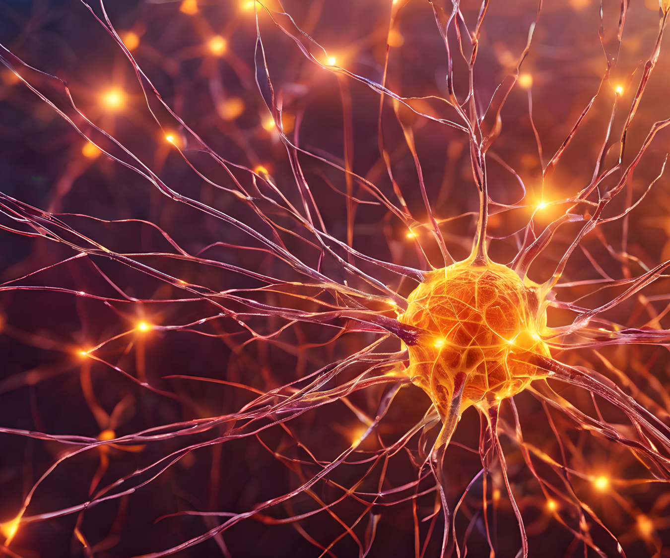
The Nervous System

The Nervous System: Structure, Function, and the Role of Neural Communication

The nervous system is one of the most important systems in the human body. It controls communication between the brain, spinal cord, muscles, organs, and tissues. It allows the body to respond to internal and external changes while coordinating movement, thoughts, emotions, and essential functions.

The NERVOUS SYSTEM works through specialized cells that transmit electrical and chemical signals. These signals allow the body to process information quickly and respond appropriately.

Understanding the nervous system is important in areas such as exercise science, health, and human performance because it plays a major role in movement, coordination, and adaptation.

Nervous Tissue

Nervous tissue is specialized tissue responsible for communication and control throughout the body.

It is found in the brain, spinal cord, and peripheral nerves.

Nervous tissue contains two primary types of cells:

  • Neurons
  • Neuroglia

These cells work together to receive information, process signals, and maintain proper nervous system function.

Unlike many other tissues, nervous tissue is designed for rapid communication.

It allows the body to react quickly to changes in the environment.

Neuroglia

Neuroglia, also called glial cells, are support cells within nervous tissue.

Although neurons are responsible for transmitting signals, neuroglia provide the environment needed for neurons to function properly.

Neuroglia perform several important roles, including:

  • Supporting neurons
  • Protecting nervous tissue
  • Maintaining chemical balance
  • Providing nutrients
  • Helping repair damaged tissue
  • Forming protective coverings around nerve fibers

Neuroglia are essential for healthy nervous system function.

Without these supporting cells, neurons would not be able to communicate effectively.

Neurons

Neurons are specialized cells that transmit information through electrical and chemical signals.

They are considered the primary communication cells of the nervous system.

A neuron receives information, processes signals, and sends messages to other neurons, muscles, or glands.

Neurons allow the body to perform many important functions, including:

  • Movement
  • Sensory awareness
  • Memory
  • Learning
  • Coordination
  • Reflex responses

Communication between neurons occurs through connections called synapses.

At these connections, chemical messengers called neurotransmitters help transfer information from one cell to another.

Neurosecretory Tissues

Neurosecretory tissues are specialized nervous tissues that produce and release hormones.

These tissues connect the nervous system with the endocrine system.

They allow the brain to influence hormone production and regulate important body functions.

Examples of processes influenced by neurosecretory tissues include:

  • Growth
  • Metabolism
  • Stress responses
  • Reproduction
  • Fluid balance

The relationship between the nervous system and endocrine system helps maintain balance within the body.

Structures of a Neuron

A neuron contains several structures that allow it to receive and transmit information.

The three main components of a neuron are:

  • Cell body
  • Axon
  • Dendrite

Each structure has a specific role in neural communication.

Cell Body

The cell body, also called the soma, is the main control center of the neuron.

It contains the nucleus and other cellular structures needed for survival.

The cell body processes information received from other neurons.

It also maintains the functions necessary for the neuron to remain active.

Dendrites

Dendrites are branch-like structures that receive signals from other neurons.

They extend outward from the cell body and increase the surface area available for communication.

When a neuron receives information, dendrites carry these signals toward the cell body for processing.

The number and structure of dendrites help determine how effectively a neuron can communicate.

Axon

The axon is a long structure that carries signals away from the cell body.

It transmits electrical impulses to other neurons, muscles, or glands.

Some axons are covered with a protective substance called myelin.

Myelin helps signals travel faster and more efficiently.

The axon allows communication over long distances within the body.

The Central Nervous System

The central nervous system (CNS) is one of the two major divisions of the nervous system.

The nervous system is separated into two main divisions:

  • Central nervous system
  • Peripheral nervous system

The central nervous system includes:

  • Brain
  • Spinal cord

The CNS acts as the main processing center of the body.

It receives information, interprets signals, and creates responses.

The Peripheral Nervous System

The peripheral nervous system includes all nerves outside of the brain and spinal cord.

Its primary role is communication between the CNS and the rest of the body.

The peripheral nervous system carries sensory information toward the CNS and sends motor commands to muscles and organs.

This division allows the brain and spinal cord to communicate with every part of the body.

Nervous System and Movement

The nervous system plays a critical role in physical movement.

When a person decides to move, the brain sends signals through the spinal cord and peripheral nerves to activate muscles.

This communication allows muscles to contract and produce movement.

Exercise training can improve nervous system efficiency.

Strength improvements, especially during early stages of training, often occur because the nervous system becomes better at activating muscles.

Importance of the Nervous System in Health and Fitness

Understanding the nervous system helps explain how the body adapts to exercise.

Fitness professionals use this knowledge to improve:

  • Movement quality
  • Coordination
  • Strength development
  • Reaction time
  • Injury prevention

The nervous system influences every movement pattern.

A healthy nervous system is essential for physical performance and daily function.

Conclusion

The NERVOUS SYSTEM is a complex communication network that controls and coordinates the body’s activities.

Nervous tissue contains neurons and neuroglia, which work together to transmit signals and support communication.

Neurons contain three main structures: the cell body, axon, and dendrite. These structures allow neurons to receive, process, and transmit information.

The nervous system is divided into the central nervous system and peripheral nervous system, allowing communication between the brain, spinal cord, and the rest of the body.

By understanding nervous system structure and function, we gain a better understanding of movement, health, and human performance.

Muscles

Understanding Muscle: Contractions, Actin, Myosin, and Sarcomere Function

Muscles are essential for movement, stability, posture, and overall physical performance. Every movement, from walking and lifting weights to breathing and maintaining balance, depends on the coordinated action of muscle tissue.

To understand how muscles create movement, it is important to understand the microscopic structures responsible for force production. Muscle contractions involve complex interactions between proteins, energy systems, and specialized structures within muscle fibers.

Key components of muscle function include contractions, myosin and actin, sarcomere length, and the z-disc.

The Structure and Function of Muscle

The human body contains three main types of muscle tissue:

  • Skeletal muscle
  • Cardiac muscle
  • Smooth muscle

Skeletal muscle is the type most associated with exercise and movement. These muscles attach to bones through tendons and create movement by contracting and producing force.

Skeletal muscles are made up of muscle fibers. Inside each muscle fiber are smaller structures called myofibrils.

Myofibrils contain repeating units called sarcomeres.

Sarcomeres are the basic functional units responsible for muscle contraction.

Understanding Muscle Contractions

A muscle contraction occurs when muscle fibers generate tension and produce force.

A contraction does not always mean that a muscle becomes shorter.

Muscles can create force in several ways.

Concentric Contractions

A concentric contraction occurs when a muscle produces force while shortening.

An example is the upward phase of a biceps curl.

The biceps muscle contracts and becomes shorter as it lifts the weight.

Eccentric Contractions

An eccentric contraction occurs when a muscle produces force while lengthening.

An example is lowering the weight during a biceps curl.

Although the muscle is lengthening, it is still actively producing tension.

Eccentric contractions are important for controlling movement and absorbing force.

Isometric Contractions

An isometric contraction occurs when a muscle produces tension without changing length.

An example is holding a plank position.

The muscles are active, but there is no visible movement at the joint.

The Role of Myosin and Actin

Muscle contraction depends on two major proteins:

  • Myosin
  • Actin

These proteins interact through a process called the sliding filament mechanism.

Actin is a thin protein filament.

Myosin is a thicker protein filament with structures called myosin heads.

During contraction, myosin heads attach to actin and pull the actin filaments inward.

This movement causes the sarcomere to shorten.

When many sarcomeres shorten together, the entire muscle produces movement.

The Sliding Filament Theory

The sliding filament theory explains how muscles generate force.

During a contraction:

  1. A signal from the nervous system reaches the muscle.
  2. Calcium is released inside the muscle fiber.
  3. Calcium allows myosin to attach to actin.
  4. Myosin pulls actin filaments closer together.
  5. The sarcomere shortens.
  6. The muscle produces force.

The actin and myosin filaments do not become shorter themselves.

Instead, they slide past each other.

This creates muscle shortening and movement.

Sarcomere Length and Muscle Performance

The length of a sarcomere affects how much force a muscle can produce.

A sarcomere must have an ideal length for maximum force production.

If a sarcomere is too stretched, actin and myosin have limited overlap.

This reduces the ability of myosin to connect with actin.

If a sarcomere is too shortened, the filaments interfere with each other.

This also reduces force production.

The best force production occurs when there is an optimal amount of overlap between actin and myosin.

This relationship is known as the length-tension relationship.

The Importance of the Z-Disc

The z-disc is a structure located at the end of each sarcomere.

It acts as an anchor point for actin filaments.

Each sarcomere is positioned between two z-discs.

When a muscle contracts, the z-discs move closer together as the sarcomere shortens.

The z-disc provides structural organization and allows force to be transferred throughout the muscle fiber.

Without the z-disc, the muscle would not be able to efficiently transmit force.

How Exercise Changes Muscle Function

Exercise creates stress on muscles.

The body responds by adapting.

Resistance training is especially effective for increasing muscle strength and size.

Strength improvements occur because of several changes:

  • Increased muscle fiber size
  • Improved nervous system communication
  • Greater ability to produce force
  • Increased efficiency of muscle contractions

During resistance training, microscopic damage occurs within muscle fibers.

The body repairs this damage and adapts by creating stronger muscle tissue.

Muscle Growth and Adaptation

Muscle growth, also called hypertrophy, occurs when muscle fibers increase in size.

This process is influenced by:

  • Training intensity
  • Exercise volume
  • Recovery
  • Nutrition
  • Hormonal responses

Consistent training provides the stimulus needed for adaptation.

However, recovery is equally important.

Muscles need time to repair and rebuild after exercise.

The Role of Energy in Muscle Contraction

Muscle contractions require energy.

The primary energy source used directly by muscles is ATP, or adenosine triphosphate.

ATP provides the energy needed for myosin heads to attach, pull, and release from actin.

The body produces ATP through different energy systems depending on the activity.

Short, powerful movements rely heavily on stored energy systems.

Longer activities rely more on aerobic energy production.

Why Understanding Muscle Function Matters

Understanding muscle anatomy and function helps fitness professionals create effective training programs.

Knowing how contractions work helps trainers select appropriate exercises.

Understanding actin, myosin, sarcomere length, and the z-disc provides insight into how muscles produce force and adapt.

This knowledge also helps explain why proper training, recovery, and progression are necessary for improvement.

Conclusion

Muscle function depends on a highly coordinated process involving contractions, protein interactions, and specialized structures.

Actin and myosin work together through the sliding filament mechanism to create force.

Sarcomere length influences how effectively muscles can contract.

The z-disc provides structure and helps transfer force throughout muscle fibers.

Through exercise and proper recovery, muscles adapt by becoming stronger and more efficient.

Understanding these processes provides the foundation for improving performance, preventing injury, and creating effective fitness programs.

Tabebordbar, M., Wang, E. T., & Wagers, A. J. (2013). Skeletal Muscle Degenerative Diseases and Strategies for Therapeutic Muscle Repair. Annual Review of Pathology: Mechanisms of Disease, 8, 441–475.

Harvard Stem Cell Research article — Skeletal Muscle Degenerative Diseases and Strategies for Therapeutic Muscle Repair